The development of a microscale implantable device by Swedish researchers opens up new possibilities for the cell-based therapy of Diabetes Treatment and other illnesses.
Collaboration and Design: Diabetes Treatment
A team from KTH Royal Institute of Technology and Karolinska Institutet developed the 3D-printed device with the goal of encapsulating insulin-producing pancreatic cells with electronic sensors. The work’s findings were published by the researchers in the journal Advanced Materials.
Through their collaboration, KTH and Karolinska Institutet have made it possible to accurately place micro-organs, such as pancreatic islets or islets of Langerhans, inside the eye without the need for sutures. It presents the prospect of using the eye as a base for cell-based therapy, for example, to treat Type 1 or Type 2 diabetes.
Diabetes Treatment
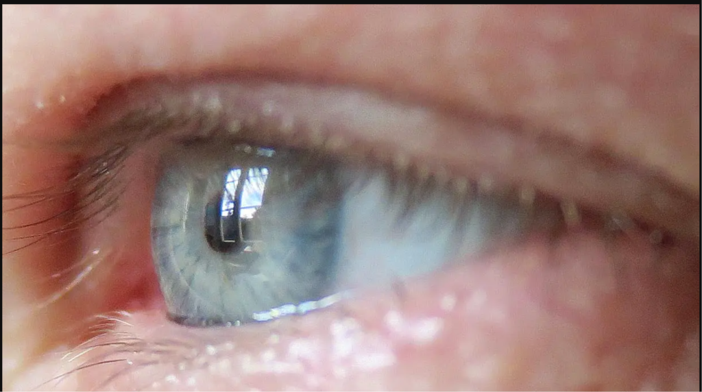
“The eye is our only window into the body, and it’s immune-privileged,” says Anna Herland, senior lecturer in the Division of Bionanotechnology at SciLifeLab at KTH Royal Institute of Technology, and the AIMES research center at KTH and Karolinska Institute. Credit: David Callahan/KTH Royal Institute of Technology
Also read : Cancer : Did Cancer Already Exist Before It Was Discovered That Man-Made Substances Might Cause It?
Advantages of the Eye
The eye is perfect for this technology, according to Anna Herland, senior lecturer in the Division of Bionanotechnology at SciLifeLab at KTH and the AIMES research centre at KTH and Karolinska Institutet, because it lacks immune cells that would react negatively during the initial stage of implantation. Because of its transparency, changes to the implant over time can be studied both visually and microscopic.
“The eye is our only window into the body, and it’s immune-privileged,” Herland says.
Diabetes Treatment
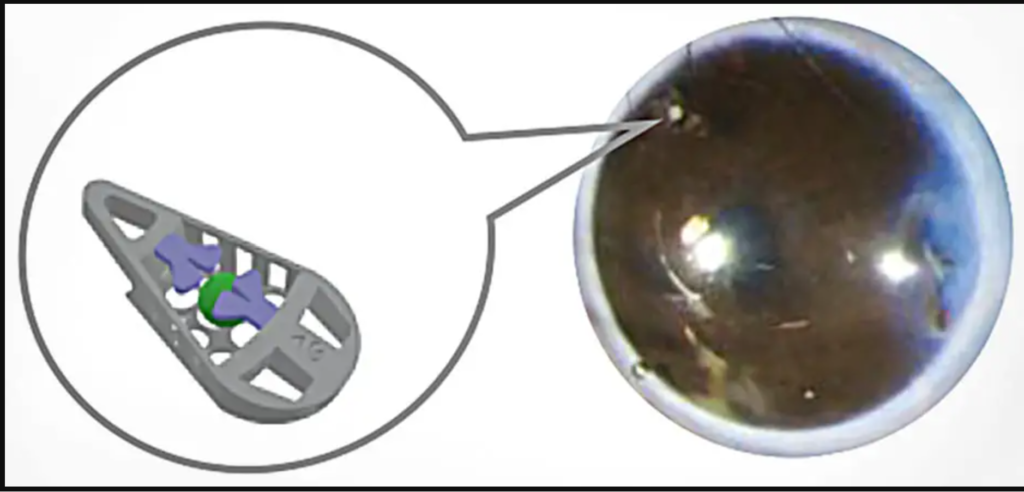
Illustration of the microdevice, and photograph of its position in the eye of a lab mouse. Credit: Hanie Kavand, Montse Visa, Martin Köhler, Wouter van der Wijngaart, Per-Olof Berggren, Anna Herland
Device Design and Functionality
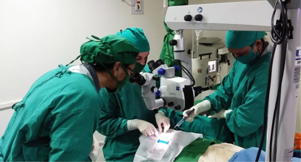
The apparatus, shaped like a wedge and measuring roughly 240 micrometres in length, enables the mechanical fixation of the structure at the anterior chamber of the eye’s (ACE) angle between the iris and cornea. The piece shows how a gadget was fixed mechanically for the first time in the anterior chamber of the eye.
Diabetes Treatment
According to Wouter van der Wijngaart, a professor in KTH’s Division of Micro- and Nanosystems, “we designed the medical device to hold living mini-organs in a micro-cage and introduced the use of a flap door technique to avoid the need for additional fixation.”
Promising Results in Animal Testing
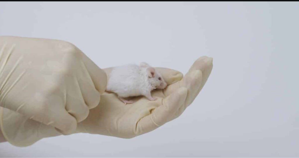
According to Herland, in mice experiments, the device remained in the living organism for several months, and the mini-organs quickly assimilated into the blood arteries of the host animal and resumed their regular functions.
Diabetes Treatment
With years of experience transplanting islets of Langerhans to the anterior chamber of the mouse eye, Per-Olof Berggren, a professor of experimental endocrinology at Karolinska Institutet, made a valuable contribution to the research.”Among other things, the current unit will serve as the foundation for our ongoing efforts to create an integrated microsystem for researching the survival and operation of the islets of Langerhans in the anterior chamber of the eye,” according to Berggren. “Transplanting Langerhans islands to the anterior chamber of the human eye is undergoing clinical trials in patients with diabetes, so this is also of great translational importance.”
Overcoming Obstacles
According to Herland, the approach gets around one barrier to the advancement of cell therapies, such as those for diabetes. Specifically, non-invasive techniques for assessing the function of the graft and directing treatment are not required to guarantee long-term transplant success.

Diabetes Treatment
“We’ve made a first step towards developing sophisticated medical microdevices that can track and identify the health of stem cells,” the researcher adds. According to her, the architecture allows for the placement of mini-organs like organoids and islets of Langerhans without compromising the cells’ ability to receive nourishment. “Future integration and use of more sophisticated device functions, like integrated electronics or drug release, will be made possible by our design.”
Reference: “3D-Printed Biohybrid Microstructures Enable Transplantation and Vascularization of Microtissues in the Anterior Chamber of the Eye” by Hanie Kavand, Montse Visa, Martin Köhler, Wouter van der Wijngaart, Per-Olof Berggren and Anna Herland, 10 October 2023, Advanced Materials.
DOI: 10.1002/adma.202306686
Also read : Researchers Determine The Reason For The Inexplicable 35 African Elephants Death In Zimbabwe


